


To date, science knows about 280 types of worms that can grow and live in the human body, parasites in various organs and tissues. The frequency of human worm infections depends on the climatic and socio-economic conditions of a particular region (in underdeveloped countries, especially in areas located in tropical and subtropical zones, the level of parasitic infections is much higher than in economically developed countries).
How humans are infected with helminths
- Biohelminthiasis (infection from animals).
- Helminthiases are contagious (transmitted from person to person).
- Geohelminthiasis (a disease caused by a parasite that runs one of its life cycles on earth).
Factors influencing the manifestations of helminthiasis
- The way parasites enter the body;
- Level of helminth adaptation to the human body;
- Population density (number) of parasitic individuals;
- Worm habitat (tissue parasite living in soft tissue thickness, and luminal living in hollow organ lumen). Some helminths in different phases have luminal and tissue forms. The degree of larval and developing worms, as a rule, causes more obvious pathological changes.
In the absence of re-infection, the number of adult parasites in the human body does not increase. This feature distinguishes helminthic invasion significantly from diseases caused by bacteria, viruses, fungi and protozoa.
Worms in humans: symptoms
Helminthiasis is a disease characterized by 2 stages of course (acute, from two weeks to two months) and chronic (from several months to several years).
Symptoms of the acute phase of helminthiasis
The first signs of the disease can appear at different times (most often after 2-3 weeks, with ascariasis - after 2-3 days, and with filariasis, the incubation period can last 6-18 months).
In the acute stage of parasitic invasion, the most typical symptom is an allergic reaction (antibodies are produced against migrating parasitic larval antigens). Often in people infected with worms, an itchy rash appears on the skin, prone to repeated travel, increased lymph nodes, general or local edema, muscle and joint pain may occur. Also, migrating parasitic larvae can cause chest pain, coughing, choking attacks, nauseous stools, nausea and vomiting.
At the same time, the acute phase of helminthiasis can be accompanied by more serious disorders (severe forms of pneumonia, hepatitis, allergic myocarditis, hepatosplenomegaly (enlargement of the liver and spleen), meningoencephalitis).
The number of eosinophils in the blood increases (eosinophilia) and the normal quantitative ratio between protein fractions is disrupted (dysproteinemia).
Signs of chronic helminthiasis
The symptomatology of the chronic phase directly depends on which organ is "inhabited" by the parasite, as well as its size and number play an important role.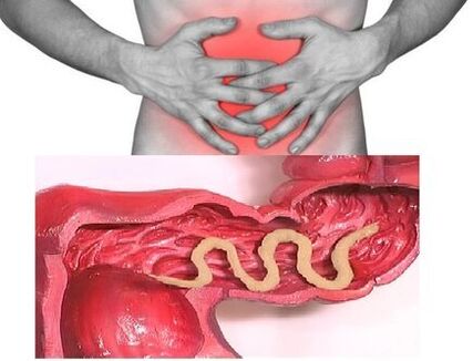 Thus, when parasites are present in the intestines of a single individual, the disease can be asymptomatic (except in cases of infection with very large parasites). The characteristic signs of the chronic phase of intestinal helminthiasis are dyspeptic disorders. In children, asthenoneurotic syndrome and pain are more pronounced. With massive roundworm attacks, there is a possibility of intestinal obstruction, obstructive jaundice and pancreatitis.
Consuming all the nutrients necessary for their vital activities from the host body, helminths cause digestive disorders, disorders of absorption of vitamins, minerals, carbohydrates, proteins and fats. At the same time, worm excretory products inhibit normal intestinal microflora and reduce immunity.
In people suffering from helminthiases, due to a weakened immune system and increased cell division (as a result of the continuous recovery of tissue damaged by parasites), the risk of malignant tumors increases significantly.
Types of parasitic helminths in the human body
The causative agents of human helminthiasis are 2 types of worms: round (nematodes) and flat (tapeworms and worms).
Round Worms
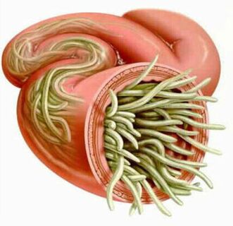
Pinworm
The parasites that cause enterobiasis are small thin cavity worms (up to 10mm) with a grayish white color. Infections occur through food (through the mouth). The reason is dirty hands. Parasite eggs can be on the ground, in the fur of infected animals, unwashed vegetables and fruits, and so on. At the same time, with enterobiasis, cases of self-infection often occur (especially in children), as a result of itching of the itchy area and subsequent swallowing of the egg. 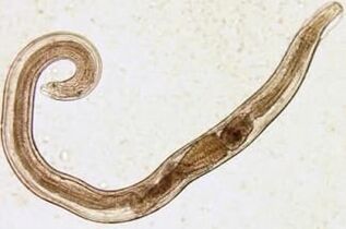 Cream worm larvae develop within two weeks in the digestive tract. Once transformed into adults, parasitic worms in the lower part of the small part and the upper part of the large intestine.
Even at the larval stage, cream worms begin to harm the body of their host, producing enzymes that irritate the intestinal wall and cause the development of inflammatory processes. Adult parasites attach to or penetrate the inner lining of the intestinal mucosa, disrupting integrity and contributing to the connection of secondary bacterial infections. If the perforation of the cream worm on the wall of the small intestine, peritonitis can develop. Also, due to irritation of intestinal receptors, motor function and secretion of the gastrointestinal tract are disrupted, leading to the formation of gastroduodenitis, enteritis, etc. In childhood, long-term enterobiasis can cause nerve disorders and delay in physical development.
Ascaris
Ascaris is a large red-yellow spider-shaped parasite, reaching 40 cm (female) and 15-25 cm (male) in adulthood. Without a suction cup or other binder, the ringworm can move freely towards the mass of food. Eggs born of parasitic females are excreted along with feces.
Infection with ascariasis occurs when mature eggs are ingested with water or vegetables and fruits that have not been washed with soil particles. Once eggs enter the intestine, mature larvae emerge from them. Then, penetrating the intestinal wall, they reach the heart through the bloodstream, and from there they enter the lungs. Through the pulmonary alveoli, the larval worm larvae through the respiratory tract re-enter the oral cavity. After repeated swallowing, the parasite reaches the small intestine, where it develops into an adult. The worm lives for 12 months, then dies and is excreted along with the feces. In the gut of one host, one and several hundred individuals can live.
In the intestinal phase of their existence, the ringworm, endowed with the ability to perform rotational movements, can penetrate the narrowest openings. These parasitic features often lead to the emergence of relatively serious complications (obstructive jaundice or pancreatitis). Allergens secreted by ringworm can cause severe allergic reactions. A large number of adults can cause intestinal obstruction, and worms that enter the airways sometimes cause shortness of breath.
Vlasoglav
Vlasoglav, the causative agent of trichocephalosis, is a white helminth parasite in the early part of the large intestine and reaches a size of 4-5 cm. Parasites feed on the blood and mucosal tissue of the rectum.
Whip eggs placed by females on the intestinal wall come out along with feces. Their development takes place in the environment (optimally on the ground). Eggs with parasitic larvae cooked in them enter the body through digestion, through dirty hands, with unwashed water or vegetables and fruits.
With a small number of worms, trichocephalosis is not symptomatic. At severe levels (with massive invasion), patients experience abdominal pain, severe diarrhea, sometimes accompanied by rectal prolapse. This condition is most often observed in weak children. With the moderate phase of trichocephalosis, growth retardation of the child is possible.
Trichinella
The causative agent of trichinosis is a small round helminth 2-5 mm long. Infection occurs while eating poorly roasted meat (pork, bear meat, wild boar).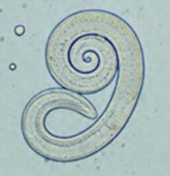 Penetrating into the intestine, parasitic larvae mature in 3-4 days into a sexually mature individual. The life span of the worm is 40 days, after which the parasite dies. By penetrating the intestinal wall, the larvae enter the bloodstream and are carried to all organs of the human body, settling in the muscles. In this case, the respiratory and facial muscles, as well as the flexor muscles of the limbs, are most often affected.
In the first few days after the invasion, the patient complains of abdominal pain. Then, after about 2 weeks, the body temperature rises to 39-40 C, an itchy rash appears on the skin, muscle pain develops, and the face swells. During this period, in the event of a major infection, there is a significant risk of death. After about a month, the patient recovered. The parasite is packaged in a spiral, after which it dies within two years.
Worms and nekators
These two parasites are similar in biological features, as well as the diseases they cause. In this case, it is customary to combine it with the common name (mine worm). Worms, reaching a length of 10-15 mm, parasitic in 12-p. usus. It should be noted that this is one of the most common, but, at the same time, parasites are rarely detected. Worm larvae enter the human body through the skin when in contact with contaminated soil. Next, entering the bloodstream, they, like ringworms, migrate to the lungs, and then, through the bronchi, along with the expectorant sputum, into the digestive tract. Ankylostoma parasitic in the intestine, attached to the intestinal wall. The parasite, which eats blood exclusively, bites the blood vessels that penetrate the mucous membrane, injecting anticoagulant components there. On average, adults can absorb 0, 05-0, 35 ml of blood per day. Therefore, the most typical symptoms of this helminthiasis are iron deficiency anemia, as well as changes in the ratio of protein breakdown (dysproteinemia).
Flatworms
Wide band
This is one of the largest helminths, with a length of 10-20 meters. The disease caused by this parasite is called diphyllobothriasis. The worm development cycle begins with freshwater fish or crustaceans. Larvae enter the human body, which is the final owner of the broad tapeworm, along with eggs or fillets of infected fish. Reaching the small intestine, the parasite attaches to its walls and grows into a mature individual in 20-25 days.
Diphyllobothriasis occurs against the background of gastrointestinal disorders and B12 deficiency anemia.
Disadvantages
The parasite that causes opisthorchiasis is a flatworm that reaches a length of 7-20 mm. It should be noted that more than 50% of cases of infection with hepatic fluke (also called cat fluke) occur in the Russian population. Parasitic larvae begin to develop after eggs enter fresh water (from snails that have swallowed them). Then they penetrate into the body of the fish (goldfish, kris goldfish, bream, roach). Human infection occurs when eating contaminated fish meat that has not undergone adequate heat treatment. Larvae of hepatic flux from the small intestine penetrate into the bile ducts and into the gallbladder, glued there with the help of two suction cups.
In the acute phase of helminthiasis, patients experience pain in the upper abdomen, increased body temperature, nausea, muscle pain, diarrhea, and skin rashes may occur. The chronic course of opisthorchiasis is indicated by symptoms of hepatitis, inflammation of the bile ducts, cholecystitis, disorders of the gastrointestinal tract, nervous disorders, weakness and increased fatigue. These parasites lead to the development of irreversible changes, and even after their expulsion, patients do not experience chronic inflammatory processes and functional disorders.
Pig and pig tapeworms
This parasite, almost identical in structure, reaches a length of 5-6 meters. Infection with teniarinhoses and teniasis occurs due to consumption of meat from infected cattle or pork by Finns (one form of intermediate helminthiasis). The worthy Finns, presented in the form of 0, 5 cm whitish bubbles, attach to the walls of the human small intestine and turn into adults in 3 months. The tape parasite, which consists of more than 2000 segments, continues to grow. In this case, the final part, which contains the egg, ruptures and moves freely along the large intestine to the anus, and then crawls out of the anus, or is released into the external environment along with the feces. The most notable symptom of helminthiasis is gastrointestinal tract disorders.
Echinococcus
For this parasite, a person is an intermediate host. Parasitic worms of the human body in the form of Finns. The last owner of echinococcus is a wolf, dog or cat. 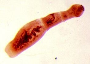 Infection occurs through food by contact with animals and with environmental objects sown with Echinococcus eggs. Upon entering the intestine, oncospheres (larvae of six hooks) develop from them. From the intestine, they enter the bloodstream and are carried throughout the body.
The worm's "favorite" parasite site is the liver and lungs. By settling on these organs, the larvae turn into Finn (echinococcal cyst), which, gradually increasing in size, begins to destroy nearby tissues. Often, echinococcosis in the diagnostic process is mistaken for a benign or malignant tumor. In addition to mechanical effects (squeezing of organs and blood vessels), rupture of echinococcal cysts occasionally occurs. This condition can cause toxic shock or the formation of some new cysts.
Alveococcus
This parasite, considered to be a type of echinococcus, is the cause of one of the most dangerous helminthiases (alveococcosis), similar to the severity of cirrhosis and liver cancer. Infection occurs when oncospheres (eggs with mature larvae) enter the intestine. There, the embryo leaves the egg and, penetrating the intestinal wall, enters the bloodstream. Furthermore, with blood flow, the parasite spreads to all tissues and organs of the body (most often it is localized in the liver). This is where the main stage of development begins in the larvae (multi-space bubbles, laurocyst formed). Each space contains the head of a parasitic embryo, which continues to grow gradually. Laurocysts are very aggressive formations that constantly grow due to enlarged bubbles, and also have the ability to grow into the liver, such as cancer metastases. Necrotic changes due to dysfunction of blood vessels undergo necrotic changes in nearby tissues. Spreading to nearby structures, alveococcus forms fibrous nodes with the inclusion of multicameral bubbles. This condition can last for several years, and therefore requires mandatory surgical intervention.
Diagnostic helminthiasis
Diagnostics of helminthic invasion include the following activities:
- taking a thorough history, helping to find out the possible cause of the infection;
- laboratory tests of stool, blood, intestinal contents 12 p, rectal and perianal mucus, muscle tissue, pulmonary sputum, bile. This analysis can reveal eggs, segments or fragments of the parasite. At the same time, an increase in eosinophil content in the blood is also a sign of helminthiasis.
- while diagnosing diseases caused by larval or tissue parasite levels, serological studies are performed (ELISA, RSK, indirect agglutination reactions, immunofluorescence analysis, etc. ).
- Ultrasound, CT and endoscopic examinations are prescribed to detect helminths affecting liver tissue.
Human worms: treatment
In the acute phase of parasitic infection, patients are given detoxification and desensitization therapy. In cases of severe disease (liver trematodes, trichinosis), glucocorticoids are used according to medical instructions.
As a specific therapeutic drug, taking into account the nature of the pathogen, a special chemotherapeutic anotelmintic agent is prescribed.
In parallel, patients are recommended to take antihistamines and enterosorbents. The final stages of treatment include the use of probiotics that normalize the intestinal microflora.
Special diets are also provided (food must be digested and low in fat).
During the period of antihelminthic therapy, patients are required to maintain strict personal hygiene (to prevent re-infection). At the same time, for many helminthiasis, all family members and people who are in constant contact with the infected must undergo treatment.
Prevention of helminthiasis
- Maintain personal and public hygiene;
- Strict adherence to cooking technology;
- Periodic checkups and preventive treatment of pets;
- Wash fresh vegetables, fruits and herbs thoroughly;
- Proper handling of river fish;
- Avoid taking raw, slightly salty and dry fish.